

What the study found
Craniofacial fibrous dysplasia osteoblasts showed signs of altered bone homeostasis, including higher proliferation, reduced osteoblastic differentiation, and a lack of mineralization ability. The study also found higher intracellular cAMP levels and a decreased osteoclastogenic potential, linked to impaired osteoclast formation in conditioned medium cultures.
Why the authors say this matters
The authors suggest that comparing craniofacial and appendicular fibrous dysplasia may help explain why lesions differ between these skeletal regions. They conclude that these features should lead to further studies on the pathogenesis of craniofacial fibrous dysplasia.
What the researchers tested
The researchers evaluated osteoblasts derived from craniofacial fibrous dysplasia lesions in vitro. They examined histological features, intracellular cAMP levels, osteoblast proliferation, osteoblastic differentiation, mineralization ability, and osteoclastogenic potential using conditioned medium cultures.
What worked and what didn't
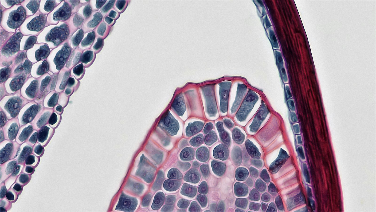
Typical histological features, including an "alphabet soup" appearance, were observed in the craniofacial lesions. The FD osteoblasts showed increased proliferation, but decreased differentiation and no mineralization ability. They also showed reduced osteoclastogenic potential because osteoclast formation was impaired in conditioned medium cultures.
What to keep in mind
The abstract does not describe sample size, controls, or detailed experimental limitations. The study focuses on craniofacial lesions and compares them conceptually with appendicular fibrous dysplasia, but it does not provide direct clinical outcomes.
Key points
- Craniofacial fibrous dysplasia osteoblasts had higher intracellular cAMP levels.
- The cells showed increased proliferation but reduced differentiation and no mineralization ability.
- Conditioned medium from these cells had reduced osteoclastogenic potential, with impaired osteoclast formation.
- Typical histological features, including an "alphabet soup" appearance, were observed in craniofacial lesions.
- The authors say the findings support further study of craniofacial fibrous dysplasia pathogenesis.
Disclosure
- Research title:
- Craniofacial fibrous dysplasia osteoblasts show altered bone homeostasis
- Publication date:
- 2026-02-24
- OpenAlex record:
- View
Get the weekly research newsletter
Stay current with peer-reviewed research without reading academic papers — one filtered digest, every Friday.