
What the study found
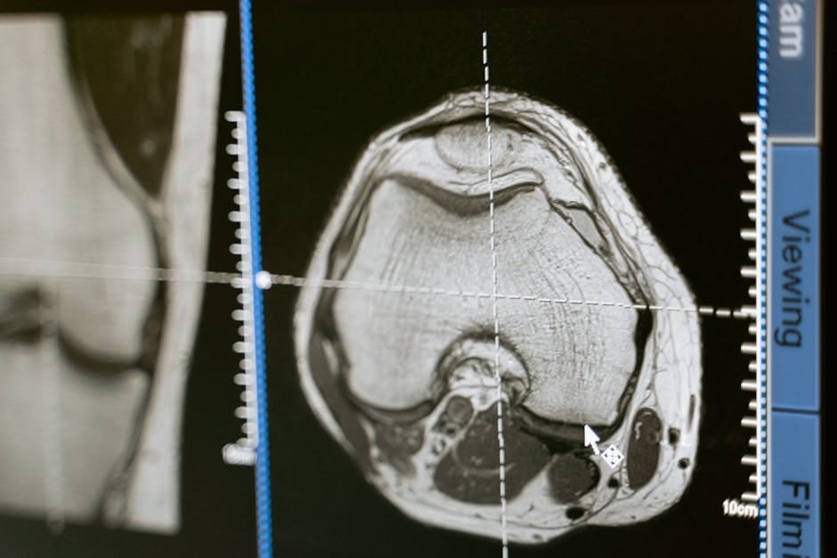
Quantitative PET/CT (positron emission tomography/computed tomography) measures of lung lesions in infected marmosets were more informative about tuberculosis treatment outcomes than bacterial burden. The lesion response profiles aligned with known clinical outcomes and could distinguish cavitary granulomas that improved from those that did not.
Why the authors say this matters
The authors conclude that combining PET/CT measures may help interpret tuberculosis treatment efficacy and better understand clinical treatment outcomes. The study suggests these lesion-level profiles can provide insight into clinical successes and failures.
What the researchers tested
The researchers measured radiographic changes in lung lesions in infected marmosets using PET/CT imaging. The animals were divided into 22 treatment arms, including monotherapies and combination drug treatments, and treated for 2 months; the team then used unsupervised clustering to combine quantitative imaging changes with terminal bacterial burden per lung lesion.
What worked and what didn't
The resulting multivariate drug response profiles aligned with known clinical outcomes. The study found that the inferior performance of the 4-month moxifloxacin-rifampicin-pyrazinamide-ethambutol regimen compared with the 6-month standard of care for people with lung cavitary TB could be predicted, and that cavitary granulomas that responded to treatment could be separated from those that failed to improve or worsened after the first month.
What to keep in mind
The abstract does not describe limitations beyond the scope of the marmoset model and the 2-month treatment period. The findings are presented for infected marmosets and are used to inform interpretation of tuberculosis treatment outcomes.
Key points
- PET/CT lesion measures were more informative about tuberculosis outcomes than bacterial burden in infected marmosets.
- Unsupervised clustering was used to build multivariate drug response profiles from imaging changes and terminal bacterial burden per lung lesion.
- The profiles aligned with known clinical outcomes and distinguished cavitary granulomas that improved from those that did not.
- The inferior performance of a 4-month moxifloxacin-rifampicin-pyrazinamide-ethambutol regimen versus 6-month standard care was predicted.
- The abstract does not list specific limitations beyond the marmoset model and 2-month treatment period.
Disclosure
- Research title:
- PET/CT lesion profiles matched tuberculosis treatment outcomes in marmosets
- Publication date:
- 2026-01-07
- OpenAlex record:
- View
- Image credit:
- Photo by MART PRODUCTION on Pexels
Get the weekly research newsletter
Stay current with peer-reviewed research without reading academic papers — one filtered digest, every Friday.